Modifying Pilates for Clients With Osteoporosis
Osteoporosis is epidemic in the United States.
Why does this concern the fitness professional? One in every 2 women and 1 in every 4 men aged 50 or older will suffer an osteoporosis-related hip, spine or wrist fracture during their lives (National Osteoporosis Foundation [NOF] 2005). Among women over 50, 1 in every 2 who walk into your exercise classes has low bone density and is at risk for fracture (NOF 2005). And research has shown that given the fragility of the osteoporotic vertebrae, most fractures are caused by the stresses of everyday life (Cummings & Melton 2002; Keller 2003). As the disease progresses, bones can become so vulnerable that fractures can occur spontaneously or through such mild trauma as opening a stuck window, lifting a light object from the floor with a rounded thoracic spine or even just coughing or sneezing.
The problem is so widespread that in October 2004, the U.S. Surgeon General released a 404-page document called Bone Heath and Osteoporosis–A Report of the Surgeon General (U.S. Department of Health and Human Services [HHS] 2004 2004). This report provides scientific information on how to improve bone health and reduce the risk of illness and injury. The document also raises the profile of a disease that too often has been overlooked.
What Is Osteoporosis?
Osteoporosis is the gradual and silent loss of bone and not a normal aging process. It is defined as a systemic skeletal disease characterized by low bone mass and microarchitectural deterioration of bone tissue, with a consequent increase in bone fragility and susceptibility to fracture (NOF 2005). Osteopenia is mildly reduced bone mass–a loss of approximately 10%-20%–indicating the onset of osteoporosis.
Clients who have their bone mineral density (BMD) tested receive a T-score, which tells them how their BMD compares with that of a young adult (25-30 years old). A standard deviation (SD) of -1 to -2.5 below the mean indicates osteopenia. An SD of more than -2.5 indicates osteoporosis. For every 1-point SD drop below the mean, fracture risk doubles.
The World Health Organization did not recognize osteoporosis as a disease until 1994. Since it is not only a newly recognized illness but also a “silent” one (bone loss stays hidden until we see its effects through changes such as height loss or kyphosis), our lack of awareness is one of its biggest allies. This lack exists even in the medical field. Studies have found that physicians too commonly fail to diagnose and treat osteoporosis, even in elderly patients who have suffered a fracture (Andrade et al. 2003; Kiebzak et al. 2002; Feldstein et al. 2003). Persons with osteoporosis usually discover bone loss insidiously. Sometimes they find that their pants are too long; or relatives notice that they are shrinking in height, they’re developing a dowager’s hump or their arms look longer. Other persons complain of waistline pain (caused when the ribs sit on the iliac crests), mid-back pain or neck pain. Hip pain is not common with osteoporosis and is present only after a fracture.
A Question of Lifestyle
In my practice as a physical therapist and a Pilates instructor, I am finding that clients and even Pilates teachers are discovering they have osteoporosis at younger and younger ages. I used to treat osteoporosis only in nursing homes, but now I’m frequently seeing osteoporotic clients in outpatient facilities, Pilates studios, and health clubs where I teach Pilates mat classes. What are we doing wrong?
Evidence suggests that lifestyle is a major culprit. Studies have found that Americans do not engage in enough physical activity, are becoming obese and do not take in enough nutrients to support good bone health (Wright et al. 2003, Gordon-Larson et al. 1999). Researchers working on the China-Oxford-Cornell Project studied rural Chinese farm families for incidence of osteoporosis, heart disease and cancer and found that even with low calcium intake the incidence of these diseases was extremely low (Hu et al. 1994; Campbell 2005). In contrast, the researchers found a positive correlation between urbanized cultures and these illnesses, which Campbell has even dubbed “diseases of affluence” (Campbell 2005).
In the U.S. we base our habits of daily living on convenience and time management. We have computers for shopping; TVs to entertain us; and microwaves, washing machines, elevators, escalators, cars and high-tech devices to “save time” and to make our lives more efficient. But more efficiency means we are increasingly sedentary, and sedentary living is bad for our bones. We walk less, we bear less weight, and we do less physical labor. In short, our high-tech labor-saving lifestyle is destroying our bones!
Two Types of Bone
Bone is alive and continuously changing, remodeling itself, storing minerals, forming blood cells and providing a strong and hopefully stable structure that can protect organs and to which muscles and ligaments can attach. There are two main types of bone, cortical and trabecular.
Cortical bone is arranged in long, parallel, compact lines and found in the long bones of the femur, tibia, humerus, radius and ulna. Trabecular, or cancellous, bone is sponge-like and found in the vertebral bodies, in the neck of the femur (hip) and at the joint surfaces of all bones. Trabecular bone has an 80% faster metabolic turnover rate than cortical bone and is affected to a much greater degree by osteoporosis. Luckily, trabecular bone makes up only 20% of our total skeletal mass, whereas cortical bone makes up the other 80%.
Two cell types are at work in our bones: osteoblasts (bone “builders”) and osteoclasts (bone “cleaners”). In the healthy skeleton these two cell types are continuously working in sync, with osteoclasts breaking down old bone and osteoblasts replacing it with new bone. In someone with osteoporosis, however, the osteoclasts continue to break down old bone, while the osteoblasts get lazy. Bone breaks down more quickly than it is remodeled, and over time, bone mass and density decrease.
While modern medicine offers significant benefits for those at risk for osteoporosis, studies clearly suggest that individuals can do a great deal to promote their own bone health. Engaging in regular weight-bearing exercise, following a bone-healthy diet and avoiding behaviors such as smoking and drinking excessive amounts of alcohol can all contribute to strong, healthy bones (HHS 2004).
The Role of Pilates
The importance of weight-bearing exercise that loads and strengthens bone cannot be underestimated. In fact, research has shown that physical exercise alone can halt the progression of bone loss (Smith & Gilligan 1987). And according to the Surgeon General’s Report, “Health and Fitness professionals can play a major role in . . . identifying and advising high-risk individuals and those who have osteoporosis” (HHS 2004).
One program that is often suggested for building strength is Pilates. For most people, this is a great idea. But, despite the media hype, is Pilates safe for clients whose bones are compromised? To teach safe and effective programs, all Pilates instructors should be educated about osteoporosis and know the precautions that apply to clients at risk for fracture. What instructors must know–if they are to help rather than harm these clients–is who is at risk and which moves are contraindicated. Without such knowledge, their clients may end up breaking a bone even as they’re exercising to build bone strength.
Which Clients Are High-Risk?
Let’s say a client in her early 50s says she has had a BMD test and was diagnosed with osteopenia, or slightly low BMD. Do the same contraindications apply to her as would apply to a client with osteoporosis?
The thoracic spine is the area of the spine at greatest risk of fracture. According to a famous study by Sinaki and Mikkelsen (1984), T7, T8 and T6 (the spinal bones just between the shoulder blades) are, in that order, the spinal bones most prone to fracture. That’s because the vertebral bodies get smaller as you move up the vertebral column and also because the thoracic vertebral bodies’ orientation toward flexion loads the spine anteriorly. However, dual-energy x-ray absorptiometry (DEXA)–commonly used to test bone density–does not view the thoracic spine because it is surrounded by ribs and the sternum, which would skew the results of the BMD report. DEXA views the lumbar spine.
Statistically, we know that bone density decreases from the cervical to the lumbar spine. However, bone size and ability to distribute force load decrease from the lumbar to the cervical spine. So if someone has osteopenia of the lumbar spine, an exercise specialist should assume that the person may have osteoporosis of the thoracic spine (Grote et al. 1995). What’s more, until the results of the client’s next bone density test are known, the instructor doesn’t know if bone loss is progressing as he or she is working with the client. Therefore, all exercise specialists should use the same precautions for clients with osteopenia as for those with osteoporosis.
Contraindications for Clients With Low Bone Density
In 1984 Sinaki & Mikkelsen tested four separate groups of subjects with osteoporosis. Group 1 performed only extension exercises (similar to Pilates Double Leg Kicks), Group 2 performed only flexion exercises (curl-ups in hook-lying position with feet on the floor; similar to the Pilates Hundred or Roll-Ups), Group 3 performed both extension and flexion exercises, and Group 4 did no exercises.
The results:
Group 1 EXTENSION: 16% had another wedge or compression fracture
Group 2 FLEXION: 89% had another wedge or compression fracture
Group 3 EXT + FLEX: 53% had another wedge or compression fracture
Group 4 NO EXERCISE: 67% had another wedge or compression fracture
The importance of these results cannot be overstated. Sinaki & Mikkelsen found that 89% of the people who performed only flexion exercises suffered additional fractures during the study. This indicates that it is harmful and dangerous to allow clients to perform flexion exercises when they have known osteoporosis! Many subsequent research papers have affirmed this (Keller 2003; Meeks 2004; Bassey 2001). Forward flexion causes excessive compression force on the anterior (or front) surface of the vertebral bodies, where most of the trabecular bone is located. In those with low bone density of the spine, the weakened bone cannot withstand such force and fractures may–or will–occur. Compression forces on the vertebrae are also excessive during side-bending of the thoracic and upper-lumbar spine. Forward flexion, side-bending and– especially–forward flexion combined with rotation are therefore contraindicated for clients with osteoporosis–and hence for clients with osteopenia.
Spinal extension is a different story. The posterior surface of the vertebral bodies contains the pars interarticularis, the pedicals and the lamina, which have a higher composition of cortical bone and are at less risk for fracture. These areas do get compressed as the spine moves into extension, but the movement is much less risky than flexion because of the strength of cortical bone. One research study showed that people with stronger back extensor muscles had higher bone density in their spines (Sinaki et al.1986). Another found that strong back extensors correlated with fewer vertebral fractures and increased bone mineral density (Sinaki et al. 1996 & 2002). The problem is that clients intuitively avoid spinal extension because of the “bone on bone” feeling at the end range during back arching. They tend to like flexion because it feels soft owing to the cushioning of the disks.
Introducing Modified Pilates
When clients with low bone density or newly healed fractures are ready to start a strengthening program, modified Pilates is an option. But safety must be paramount. The risk of additional fracture within 1 year in clients who have already experienced at least one vertebral fracture is 500% (Lindsay et al. 2001)! For maximum safety and benefit, it is crucial to follow these steps in this order:
1. Make Sure Clients Have Obtained a Physician’s Clearance to Do Pilates. Anyone who has osteoporosis or is at high risk for it must have clearance before beginning a Pilates program.
2. Use Safe Evaluation Techniques. Do not test the spine’s mobility! The following tests are safe to use:
- Functional Reach Test: Assess how far the client can reach forward without excessive rounding of the thoracic spine. Normal is about 10-12 inches.
- Lifting: Have the client attempt to lift 5-10 pounds from the floor using proper knee and spine alignment.
- Supine to Sit using “log rolling” method.
- Sit to Stand without using hands and with knees apart.
- Hip Hinge: Check to see if the client can flex at the hip joint without rounding the low back.
- Abdominal Strength Test–Leg Lowering: The head is on the floor, or if the client is kyphotic, on a small pillow. The lumbar spine should be flat. Have the client perform single-leg lowering first, and then double-leg lowering if this is possible with a flat back.
- Balance Test–Tandem Stand: Instructor must closely guard the client. Test a 10-second stance on one leg, or single-leg heel-raises 10x.
3. Protect the Spine From Fracture. Before clients begin a program, make sure they clearly understand which moves are contraindicated and which are protective. Most important, teach them to avoid all flexion, side-bending and rotation (with osteoporosis and osteopenia of the spine) (Bonner 2003; Meeks 2004).
4. Teach Clients Their Neutral or Optimal Spine Position. For some clients, neutral spine will be impossible, in which case teach them their optimal spinal position (as near neutral as possible).
5. Teach the L-Shaped Hip Hinge. Use the hip hinge to instruct clients how to disassociate spine movement from hip movement.
6. Teach Proper Breathing. Teach costal breathing, in which the ribs expand posterolaterally (bucket-handle–style, encouraging breathing into the lower back) and the transversus abdominis muscles are contracted to prevent abdominal expansion or bulging. Placing a strap around the lower ribs at or near the level of the xyphoid process will give clients feedback. They should be able to expand 1½-2 inches. Teach them to avoid lifting the chest wall or flaring the ribs.
6. When All of the Above Are Mastered, Then Begin the Bone-Building Program. Progress clients safely (see below for details)–and remember, exercise should still be fun!
Note: Do not allow clients to exercise to the point of caloric drain. Evidence has shown that women who exercised up to 5 hours per week had increased BMD, whereas women who exercised more than that showed a decrease in BMD. The conclusion was that exercise to the point of caloric drain or amenorrhea is associated with stress fractures and osteoporosis (Michel, Bloch & Fries 1989).
The Bone-Building Program
General principles:
- Throughout the program, emphasize body awareness, axial elongation (see “Case Study: Ilda” on page TK)alignment (Meeks 2004; Bonner 2003).
- Focus especially on the abdominals–but without using “crunches,” which place the spine at risk for fracture. The 90/90 Posterior Tilt and the 90/90 Lower Abs Leg Lowering exercises (see page TK) and are effective for abdominal strengthening.
- Require clients to maintain their neutral or optimal spine position during most exercises.
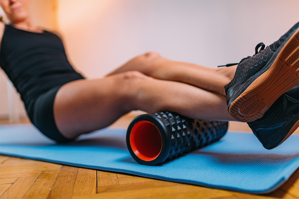
- Encourage clients to improve their thoracic spine extension (through backward bending, which is safe for the spinal vertebrae) to prevent or reduce thoracic kyphosis (dowager’s hump). A safe Foam Roller exercise that can be helpful for this purpose is the Thoracic Spine Extension.
Osteoporosis Statistics
Today, 10 million Americans have osteoporosis, and an additional 34 million have osteopenia, placing them at increased risk for osteoporosis (NOF 2005).
- Each year more than 1.5 million Americans suffer an osteoporosis-related fracture (NOF 2005).
- One in 2 American women and 1 in 4 American men over age 50 will suffer an osteoporosis-related fracture in her or his lifetime (NOF 2005).
- Among hip fracture patients, 1 in every 5 ends up permanently in a nursing home (Salkeld et al. 2000)
- People on bed rest lose about 1% of their bone mineral density per week (Smith & Gilligan 1987).
Sidebar: Risk Factors for Bone Loss?
Maybe we can understand the increasing rates of osteoporosis and fractures in America when we stop to consider just how many risk factors there are for bone loss! Sedentary living and calcium deficiency are two major culprits, but there are many other causes.
Types of diseases that increase one’s risk of osteoporosis include genetic disorders, hypogonadal states, endocrine (including thyroid) disorders, gastrointestinal diseases, hematologic disorders, rheumatic and autoimmune diseases, alcoholism, congestive heart failure, depression, emphysema, end-stage renal disease, epilepsy, idiopathic scoliosis and multiple sclerosis. Other risk factors are as follows:
Unavoidable Risk Factors
Caucasian, Northern European or Asian ethnicity
family history
menopause
phenotype: small, fine bonesBone Loss Accelerators
insufficient vitamin D
insufficient calciumBone Depleters
caffeine
protein
fiber
acidifying foods
salt
alcohol
physical inactivity
smokingWomen’s Health Issues
late menarche
irregular menstrual cycles
exercise-induced amenorrhea
previous or early hysterectomy
extended lactationEating Disorders
anorexia
bulimiaMedications That Cause Secondary Osteoporosis
anticoagulants (e.g., heparin)
anticonvulsants
cyclosporin and tacrolimus
chemotherapeutic drugs
gonadotropin-releasing hormone agonists
lithium
methotrexate
parenteral (i.e., intravenous) nutrition
antacids with aluminum
thyroid hormone therapy (thyroxine)
corticosteroidsRisk Factors for Fracture
non-Black ethnicity
female relatives with osteoporosis
early menopause (before age 45)
short and thin frame
amenorrhea
irregular periods
nulliparity (childlessness)
scoliosis
kyphosis (humpback or rounded shoulders)
height loss (more than 1 inch or 3 centimeters)
lactose intolerance
impaired vision
neurological impairment
balance impairment
history of fractures
high alcohol use (more than 1 drink per day)
high caffeine use (more than 1-2 cups per day)
smoking
low-calcium diet
lack of vitamin D
high-salt diet
high-protein diet
chronic diarrhea, surgical removal of stomach/small intestine or Crohn’s Disease
daily use of cortisone (prednisone, glucocorticoids) for more than 3 months
use of these medications:
- drugs that sedate or alter balance (they increase risk of fracture 2-3 times)
- sleep medications
- thyroid medication (over 2 grains), Synthroid, Lavoxil,
- phenytoin (Dilantin)
- aluminum-containing antacids
- long-acting benzodiazepines (e.g., Valium®)
- Depo-Provera
- Lupron
Clients with 3-5 risk factors should have their bone mineral density tested (HHS 2004; Foundation for Osteoporosis Research and Education 2002).
Sidebar: What’s Safe and What’s Not
Safe Pilates Mat Exercises
Hundred–with head down
Single Leg Circles
Single Leg Stretch–head down
Double Leg Stretch–head down
Single Leg Stretch With Straight Legs–head down
Double Leg Stretch With Straight Legs/Lower Lift–head down
Criss-Cross–head down
Swan-Dive (1 only)
Single Leg Kick
Double Leg Kick
Shoulder Bridge–not too high
Side Kick
Hip Circle/Hip Twist With Stretched Arms–neutral spine
Swimming
Leg-Pull–Front
Leg-Pull
Side Kick Kneeling–neutral spine
Side Support–neutral spine
Push-UpContraindicated Pilates Mat Exercises
Hundred–unmodified
Roll-Up
Roll-Over–both ways
Rolling Back/Rolling Like a Ball
Scissors
Spine Stretch
Rocker With Open Legs/Open Leg Rocker
Corkscrew
Saw
Neck Pull
Scissors
Bicycle
Spine Twist
Jack-Knife
Teaser
Boomerang
Seal
Crab
Rocking
Control BalanceOther Important Exercises to Add to the Osteoporosis Exercise Program
Psoas Stretches–adapted from Eve’s Lunge on the Reformer
90/90 Posterior Tilt–modified Hundred
90/90 Lower-Abs Leg Lowering–modified Hundred, lumbar spine flat
Dead Bug–knees straight, lumbar spine flat
Prone Hip Lift–modified from Single Leg Kick
Squat/Plié Variations–done in front of the Wunda Chair, not as mat work
Standing Balance Series
Half Kneeling–“marriage proposal” position
Prone Trunk Extension With Varied Arm Positions
Modified Wall Push-Up
Foam Roller
Thoracic Spine Extension
Prone Spine Extension–modified from Trapeze Table “Swan”
Vigorous walking program
Source for Pilates exercises: Pilates, J.H., & Miller, W.J. 1945. Return to Life Through Contrology. Miami: Pilates Method Alliance.
Sidebar: Case Study: Ilda
Ilda, an 83-year-old client, presented ambulating with a 3-point cane complaining of “pain in her hip so much that it hurt to drive her car.” It turned out that she had lost 7 inches of height, dropping from 5 feet 6 inches to 4 feet 11 inches, and her lowest ribs were resting on her iliac crests. With Pilates axial elongation exercises and gentle tractioning, Ilda reported complete pain relief and even reported decreased frequency of trips to the bathroom at night. At the end of sessions, her height increased to 5 feet owing to a decrease in thoracic kyphosis.
Note: Performing tractioning on clients falls outside the scope of practice for exercise instructors. Tractioning is a physical therapy treatment. However, clients can safely perform self-tractioning.
Sidebar: Case Study: Elizabeth
Elizabeth’s Story: Elizabeth, a 36-year-old client, presented with chronic low-back pain, which had been going on for 7 years since she experienced a vertebral fracture while holding her 4-month-old baby. She reported that she sneezed while holding the baby and felt a sharp pain in her mid to low back. She tried to ignore the pain that evening but woke the next morning with severe back pain and went to an orthopedic surgeon. Without taking an x-ray, the surgeon prescribed muscle relaxants for “muscle strain” and sent her home. Since she was breast-feeding, she decided not to take the muscle relaxants.
Still in pain, Elizabeth sought help from a local chiropractor, who also did not take x-rays but did perform a high-velocity manipulation on her spine, after which she reported that she was unable to move. She called her husband, who took her to the emergency room, where x-rays revealed a compression fracture at T-12 and severe osteoporosis. Later, DEXA bone mineral density reports would show T-scores in the -4.0 range or 40% bone loss.
After further testing Elizabeth discovered that she had Grave’s disease, a thyroid disorder, and not postpartum depression as she had suspected. Her doctors advised her to stop all activities and “do nothing” for exercise except walking, saying that she had the bones of a 90-year-old or worse.
After 7 years this young woman fearfully began a modified Pilates program to re-educate her body awareness, strengthen her core and develop her thoracolumbar paraspinal muscles. As a result of the program, her endurance improved, her pain decreased and she gained more confidence in doing activities without risking further fractures.
Sidebar: Resources
Betz, S. 1999. The Osteoporosis Exercise Book. Osteo Physical Therapy.
Bonner, F.J., et al. 2003. Health professional’s guide to rehabilitation of the patient with osteoporosis. Osteoporosis International, 14 (Suppl. 2), S1-22. (Includes guidelines for designing safe exercise programs.)
National Osteoporosis Foundation, www.nof.org.
Pilates Method Alliance, www.pilatesmethodalliance.org. U.S. Department of Health and Human Services. 2004. Bone Heath and Osteoporosis–A Report of the Surgeon General. www.surgeongeneral.gov/library/bonehealth/.
Sherri Betz, PT, has been a physical therapist since 1991. A Polestar principal educator/examiner and a Gyrotonic®/Gyrokinesis™ instructor, she serves on the board of directors for the Pilates Method Alliance. She is the author of The Osteoporosis Exercise Book (Osteo Physical Therapy 1999) and creator of modified Pilates videos for special populations. She is pioneering research in Pilates for osteoporosis and geriatrics. Sherri owns TheraPilates® Physical Therapy and Gyrotonic Clinic in Santa Cruz, California.
Copyright 2005 by IDEA Health & Fitness Inc. All rights reserved. Reproduction without permission is strictly prohibited.
References
Andrade, S.E., et al. 2003. Low frequency of treatment of osteoporosis among postmenopausal women following a fracture. Archives of Internal Medicine, 163 (17), 2052-57.
Bassey, E.J. 2001. Exercise for prevention of osteoporotic fracture. Age and Aging, 30 (Suppl. 4), 29-31.
Bonner, F.J., et al. 2003. Health professional’s guide to rehabilitation of the patient with osteoporosis. Osteoporosis International, 14 (Suppl. 2), S1-22.
Campbell, T.C. 2005. The China Study. Dallas: Benbella Books.
Cummings, S.R., & Melton, L.J., 3rd. 2002. Epidemiology and outcomes of osteoporotic fractures. Lancet, 359 (9319), 1761-67.
Feldstein, A.C., et al. 2003. Older women with fractures: Patients falling through the cracks of guideline-recommended osteoporosis screening and treatment. Journal of Bone and Joint Surgery (American), 85-A (12), 2294-302.
Foundation for Osteoporosis Research and Education (FORE). 2002. Guidelines for the Physician (4th ed.). FORE.
Gordon-Larson, P., McMurray, R.G., & Popkin, B.M. 1999. Adolescent physical activity and inactivity vary by ethnicity: The National Longitudinal Study of Adolescent Health. Journal of Pediatrics, 135 (3), 301-6.
Grote, H.J., et al. 1995. Intervertebral variation in trabecular microarchitecture throughout the normal spine in relation to age. Bone, 116 (3), 301-8.
Hu J.F., et al. 1994. Bone density and lifestyle characteristics in premenopausal and postmenopausal Chinese women (part of the China-Cornell Project). Osteoporosis International, 4 (6), 288-97.
Keller, T.S., et al. 2003. Prediction of spinal deformity. Spine, 28 (5), 455-62.
Kiebzak, G.M., et al. 2002. Undertreatment of osteoporosis in men with hip fracture. Archives of Internal Medicine, 162 (19), 2217-22.
Lindsay, R., et al. 2001. Risk of new vertebral fracture in the year following a fracture. Journal of the American Medical Association, 285 (3), 320-3.
Meeks, S. 2004. The role of the physical therapist in the recognition, assessment and exercise intervention in persons with, or at risk for, osteoporosis. Topics in Geriatric Rehabilitation (October).
Michel B.A., Bloch, D.A., & Fries, J.F. 1989. Weight-bearing exercise, overexercise, and lumbar bone density over age 50 years. Archives of Internal Medicine, 149 (10), 2325-29.
National Osteoporosis Foundation (NOF). 2005. Fast facts. www.nof.org/osteoporosis/diseasefacts.htm; retrieved February 3, 2005.
Pilates, J.H., & Miller, W.J. 1945. Return to Life Through Contrology. Miami: Pilates Method Alliance.
Riggs, B.L., & Melton, L.J., 3rd. 1995. The worldwide problem of osteoporosis: Insights afforded by epidemiology. Bone, 17 (5 Suppl), 505S-11S.
Salkeld, G, et al. 2000. Quality of life related to fear of falling and hip fracture in older women: A time trade-off study. British Medical Journal, 320 (7231), 341-6.
Sinaki, M., et al. 1986. Relationship between bone mineral density of spine and strength of back extensors in healthy postmenopausal women. Mayo Clinic Proceedings, 61 (2), 116-22.
Sinaki, M, et al. 2002. Stronger back muscles reduce the incidence of vertebral fractures: A prospective 10 year follow-up of postmenopausal women. Bone, 30 (6), 836-41.
Sinaki, M., & Mikkelsen, B.A. 1984. Postmenopausal spinal osteoporosis: Flexion versus extension exercises. Archives of Physical Medicine and Rehabilitation, 65 (10), 593-96.
Sinaki, M., et al. 1996. Can strong back extensors prevent vertebral fractures in women with osteoporosis? Mayo Clinic Proceedings, 71 (10), 951-56.
Smith, E.L., & Gilligan, C. 1991. Physical activity effects on bone metabolism. Calcified Tissue International, 49 (Suppl.), S50-4.
U.S. Department of Health and Human Services (HHS). Office of the Surgeon General. 2004. Bone Health and Osteoporosis–A Report of the Surgeon General. Rockville, MD.
Wright, J.D., et al. 2003. Dietary intakes of ten key nutrients for public health, United States: 1999-2000. Advance Data (334), 104. Hyattsville, MD: National Center on Health Statistics.