Harnessing Body Scan Technology in Fitness
Science and Application for Fitness Professionals
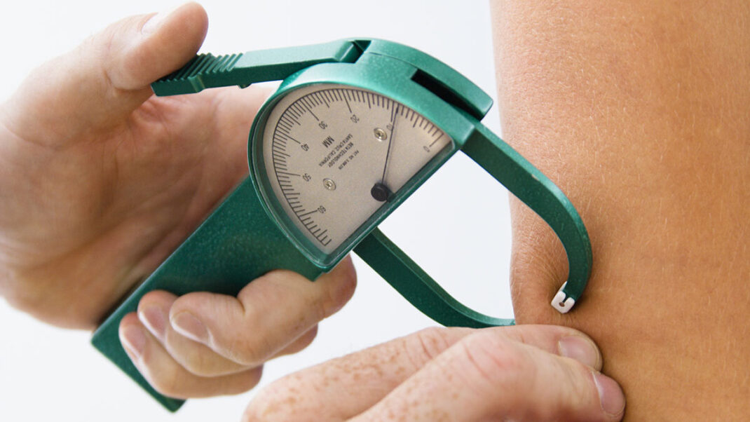
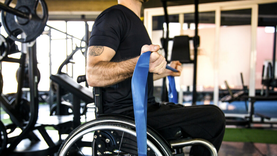
In modern fitness and wellness practice, precision and personalization are cornerstones of effective client programming. While traditional methods such as calipers, tape measurements, and scales have long served as tools for tracking progress, the integration of body scan technology offers fitness professionals a level of detail and objectivity that was previously the domain of clinical or research settings.
As fitness clients increasingly seek data-driven guidance and personalized interventions, body scan technologies provide rich, nuanced insight into body composition, health risks, and progress that can inform both training and nutrition strategies. However, to leverage these technologies effectively, fitness professionals must understand not only how the tools work, but also how to interpret and apply the data responsibly within their scope of practice.
This article explores the science behind body scan technology, its practical applications in tracking fitness and health markers, and the strategies professionals can use to set evidence-based, individualized goals using scan data.
Understanding Body Scan Technology: The Science of Assessment
What is Body Scan Technology?
Body scan technology refers to a range of non-invasive assessment methods designed to evaluate body composition and distribution of mass across different regions of the body. These technologies provide data on the proportions of lean mass, fat mass, bone density, and sometimes hydration status. Modern body scan devices typically use one or more of the following scientific principles:
- Bioelectrical Impedance Analysis (BIA):
BIA operates by passing a small, safe electrical current through the body and measuring the resistance (impedance) encountered by the current as it travels through tissues. Because lean tissue contains more water and electrolytes than fat tissue, it conducts electricity more easily. The resistance is used to estimate body water content, from which fat-free mass and fat mass can be derived (Kyle et al., 2004). Multi-frequency BIA can also provide segmental analysis (e.g., limb vs. trunk) and insights into intracellular vs. extracellular fluid. - Dual-Energy X-ray Absorptiometry (DXA):
Originally developed for assessing bone mineral density, DXA uses low-dose X-rays at two energy levels to differentiate between bone mass, lean soft tissue, and fat tissue. DXA scans provide detailed regional data, including visceral adipose tissue estimates, which are highly relevant for assessing cardiometabolic risk (Nana et al., 2015). - 3D Optical Scanning:
This method employs optical sensors or cameras to create a detailed three-dimensional image of the body’s surface. From this model, the system estimates circumferences, volumes, and, through algorithms, body composition. While indirect, 3D optical scanning offers excellent reproducibility for tracking external measurements and body shape changes over time (Esco et al., 2019).
Each method has unique strengths: BIA offers portability and affordability; DXA provides high precision and the ability to assess bone health; and 3D scanning excels at monitoring changes in body shape and posture.

Scientific Validity and Reliability
The utility of body scan technology rests on its accuracy (validity) and consistency (reliability).
- BIA is considered reliable for estimating total body water and tracking fat-free mass over time when hydration status is controlled. However, its accuracy can fluctuate with fluid shifts, food intake, exercise, and temperature (Kyle et al., 2004). Multi-frequency and segmental BIA devices improve upon earlier models, offering greater detail and accuracy, though still influenced by hydration.
- DXA is widely regarded as a gold standard for body composition analysis in both research and clinical settings. It offers precise regional breakdowns of fat and lean mass and provides bone mineral content data. DXA’s ability to estimate visceral fat is particularly valuable for assessing health risks (Nana et al., 2015). However, cost, access, and exposure to low-level radiation limit its frequent use in some fitness settings.
- 3D optical scanning shows high test-retest reliability for body shape and circumference measurements, making it an excellent tool for tracking external changes in body dimensions. Though less precise in estimating fat and lean mass than DXA, it provides actionable insights into symmetry, posture, and girth changes (Esco et al., 2019).
It is crucial for fitness professionals to recognize that no scan method is perfect, and all should be interpreted as part of a comprehensive assessment strategy.
Applications for Tracking Fitness Progress
Monitoring Body Composition Changes Over Time
One of the most valuable applications of body scan technology is the ability to track body composition over weeks, months, and years with greater granularity than weight scales alone. Body scans can reveal:
- Changes in lean body mass: Monitoring gains in muscle mass, particularly in specific regions like arms, legs, or trunk.
- Fat mass reduction patterns: Identifying whether fat loss is generalized or concentrated in particular areas (e.g., trunk or visceral regions).
- Segmental asymmetries: Detecting imbalances in muscle development that may inform programming adjustments for injury prevention or aesthetic goals.
For example, a client focused on hypertrophy might see evidence of lean mass gains even if overall weight remains constant—a sign of favorable body recomposition. Similarly, scans can show fat loss trends that might not be evident from scale weight, providing clients with more meaningful feedback on their efforts.
Supporting Client Motivation and Retention
Quantitative feedback from body scans can play a vital role in maintaining client motivation and enhancing program adherence. Unlike weight alone—which can fluctuate due to hydration or gastrointestinal contents—body composition data allows clients to see the direct impact of training and nutrition on their body’s internal makeup.
Research suggests that clients who receive detailed, objective feedback are more likely to stay committed to their fitness journey (Peterson et al., 2011). Body scan data:
- Reinforces positive behaviors, especially when scale weight stalls but body composition improves.
- Provides tangible evidence of progress that might not be visible to the naked eye.
- Helps clients appreciate the value of strength gains and fat loss beyond superficial metrics.
By focusing on these meaningful indicators of change, fitness professionals can help shift client mindset from scale-obsessed to health- and performance-oriented.

Fine-Tuning Training and Nutrition Programs
Body scan data can help fitness professionals make data-informed decisions about program adjustments. For example:
- If a client is losing lean mass during a fat loss phase, the trainer might modify resistance training intensity, volume, or dietary protein intake.
- If asymmetries in muscle mass are detected, unilateral exercises or corrective strategies may be introduced.
- If trunk fat or visceral fat is not responding as expected, a reassessment of training intensity, aerobic work, or nutritional strategies might be warranted.
This level of detail allows programs to evolve responsively, helping clients avoid plateaus and achieve better outcomes.
Applications for Assessing Health Risks
Visceral Adiposity and Cardiometabolic Risk
Modern body scan technologies, particularly DXA, provide estimates of visceral fat volume. Visceral fat—located deep within the abdominal cavity—is a potent risk factor for insulin resistance, type 2 diabetes, cardiovascular disease, and metabolic syndrome (Després, 2012).
For fitness professionals, monitoring visceral fat trends:
- Allows early identification of clients who may benefit from medical evaluation.
- Helps prioritize interventions that emphasize central fat loss, such as combined aerobic and resistance training.
- Provides quantifiable markers to show clients how training reduces health risks over time.
Emphasizing visceral fat reduction, rather than just subcutaneous fat or scale weight, can reinforce the importance of sustainable, health-focused behavior change.
Bone Health Monitoring
Body scans that assess bone mineral density (e.g., DXA) provide valuable information, particularly for populations at risk for low bone mass, such as postmenopausal women, older adults, and individuals with low body weight or eating disorders.
While fitness professionals do not diagnose conditions like osteoporosis, awareness of low or declining BMD can:
- Inform exercise prescription with a greater emphasis on weight-bearing and resistance activities that promote bone health.
- Encourage collaboration with healthcare providers for further evaluation and intervention when appropriate (Nana et al., 2015).
This adds another dimension of health support within the fitness professional’s scope of practice.

Using Scan Data for Goal Setting
Crafting SMART Goals from Scan Results
Body scan results provide a robust foundation for setting specific, measurable, achievable, relevant, and time-bound (SMART) goals. Data-driven targets might include:
- “Decrease trunk fat mass by 1.5 kg over 10 weeks while preserving lean mass.”
- “Increase lower body lean mass by 0.7 kg in 12 weeks to address strength asymmetry.”
- “Reduce estimated visceral fat by 10% over 16 weeks through combined training modalities.”
Such precision helps clients focus on realistic, evidence-based objectives, improving engagement and outcomes.
Avoiding Misuse and Misinterpretation
Fitness professionals must help clients interpret scan results responsibly. Key principles include:
- Focus on trends, not single data points: Normal biological fluctuations can cause small variations between scans. Emphasize long-term patterns rather than minor changes.
- Place body composition within a broader health context: Stress the importance of strength, mobility, cardiovascular fitness, and well-being alongside body composition.
- Educate about variability: Explain how hydration, food intake, menstrual cycle, or recent exercise can affect readings, reducing undue anxiety over minor shifts.
By fostering informed, balanced perspectives, professionals can keep clients motivated and focused on sustainable progress.
Best Practices for Implementing Body Scan Technology
Standardizing Assessment Conditions
For consistent, accurate tracking:
- Schedule scans at the same time of day, ideally in a fasted state or under controlled pre-assessment conditions.
- Encourage clients to avoid caffeine, alcohol, intense exercise, or large meals several hours before scanning.
- Promote consistent hydration practices, especially when using BIA.
Such practices minimize confounding variables and enhance data reliability.
Complementing Other Assessment Tools
Body scan technology should be integrated with:
- Performance metrics: Strength, endurance, flexibility, balance.
- Movement quality assessments: Functional screens or postural analysis.
- Subjective feedback: Energy levels, sleep quality, mood, stress levels.
This holistic approach ensures that training decisions are informed by the full spectrum of client data—not body composition alone.
Ethical Use and Communication
As with any assessment tool:
- Maintain strict confidentiality of client data and respect privacy.
- Use scan data to promote health, function, and realistic body image, not unattainable or unhealthy standards.
- Communicate results in a supportive, educational manner, empowering clients rather than inducing anxiety or discouragement.
Modern body scan technology provides fitness professionals with unprecedented insight into client physiology, offering a powerful means to track progress, assess health markers, and set tailored goals. When used thoughtfully and ethically, these tools can elevate the standard of care, foster deeper client trust, and support long-term success. The key lies in pairing the technology’s precision with professional wisdom, compassion, and a commitment to whole-person health.
References
• Després, J.-P. (2012). Body fat distribution and risk of cardiovascular disease: An update. Circulation, 126(10), 1301–1313.
• Esco, M. R., Snarr, R. L., Leatherwood, M. D., Chamberlain, N. A., Redding, M. L., McIntosh, S. E., & Williford, H. N. (2019). Comparison of total and segmental body composition using DXA and a multi-frequency bioelectrical impedance device in physically active women. Journal of Strength and Conditioning Research, 33(6), 1667–1673.
• Kyle, U. G., Bosaeus, I., De Lorenzo, A. D., Deurenberg, P., Elia, M., Gómez, J. M., Heitmann, B. L., Kent-Smith, L., Melchior, J.-C., Pirlich, M., Scharfetter, H., Schols, A. M. W. J., & Pichard, C. (2004). Bioelectrical impedance analysis—Part I: Review of principles and methods. Clinical Nutrition, 23(5), 1226–1243.
• Nana, A., Slater, G. J., Hopkins, W. G., & Burke, L. M. (2015). Effects of daily activities on dual-energy X-ray absorptiometry measurements of body composition in active people. Medicine & Science in Sports & Exercise, 47(4), 720–730.
• Peterson, M. D., Rhea, M. R., & Alvar, B. A. (2011). Applications of the dose–response for muscular strength development: A review of meta-analytic efficacy and reliability for designing training prescription. Journal of Strength and Conditioning Research, 19(4), 950–958.