Functional Anatomy: From Theory to Practice
Anatomy. Many fitness professionals would rather read about something else. They know they “have to learn this stuff,” but the thought of studying the origin, insertion point and action of each muscle fills them with dread. And it just doesn’t seem that important when helping clients lose weight, build muscle or improve their function and/or performance. However, nothing could be further from the truth. The problem isn’t the actual study of anatomy—it’s the way in which anatomy has traditionally been taught.
Most of the available information on anatomy to date has been gleaned from studying and dissecting cadavers to see where muscles attach (Gray 1995). The findings yielded the framework for the most widely held views regarding muscle function and anatomy education—muscle A goes from bone 1 to bone 2, and when the muscle contracts, it pulls these two bones together. A typical anatomy text might explain, for example, that the quadriceps muscles are responsible for extending or straightening the knee (Golding & Golding 2003). Personal trainers teach clients the leg extension exercise in order to strengthen their quadriceps, and this would seem like a valid rationale. However, in “real life” the body functions very differently.
In real life, another function of the quadriceps is to slow down knee flexion, as happens when we walk, squat or lunge (Price 2008). With this other function, we see how the quadriceps perform when a person is on two feet (not sitting on a machine), with gravity and ground reaction forces coming into play. If, as a fitness professional, you can start to think about anatomy in this alternate way and teach people how their bodies work in everyday life, clients might be more likely to pay attention to the way they move, both in and out of the gym. This increased body awareness might also improve exercise adherence. And armed with knowledge of how muscles mesh with movement, you will design more effective strategies for building muscle, burning fat, improving performance and function, and eliminating pain.
Gravity
To better understand the real-life function of muscles, it’s important to understand the role of gravity. Gravity is a constant force of attraction, put out by the earth, that pulls everything on the surface of the planet toward its core (Holzner 2006). The body must work 24 hours a day against this pull to maintain its form. If we couldn’t do this, we’d never be able to stand upright and would be crushed like aluminum cans against the ground. The skeletal system, which provides an unyielding frame for the body, enables us to maintain our structure. The function of most muscles is to limit unnecessary stress from the force of gravity and the transfer of energy (ground reaction forces) throughout the body (Price 2008). Muscles lengthen in order to slow the rate at which body parts move toward and away from each other. This lengthening action also helps maintain balance and decreases stress to joints. For example, if you were standing upright in perfect structural alignment, the skeletal system (along with ligaments) would keep you erect. In essence, your center of gravity would be balanced over a solid foundation and your muscles would not need to work much against gravity. However, if you then leaned over to pick up a pencil, your center of gravity would shift forward and the muscles on the back of your torso would have to lengthen to slow down your trunk. Otherwise you’d fall flat on your face.
Ground Reaction Forces
Muscles must also constantly deal with ground reaction forces, or impact. Newton’s third law of motion states that “for every action there is an equal and opposite reaction” (Chasan 2002). When the body—via the foot—applies force to the ground, the ground returns the same amount of force to the body—via the foot—in the opposite direction (i.e., stomping your foot transfers force down through the bottom of your foot to the ground, and ground reaction forces return the same amount of force).
What this means for the musculoskeletal system is that, in addition to dealing with the constant pull of gravity, muscles must contend with the energy that is generated and transferred into the body every time any part of it comes in contact with the ground. As with gravity, muscles do this by lengthening in order to slow down energy as it travels through the muscles and soft-tissue structures to the joints and underlying skeletal system. Absorption of ground reaction forces by the lengthening muscles also enables movement to continue, because the energy stored in the elongated muscles is then used to provide power to the muscle fibers when they subsequently contract.
For example, imagine that one end of a bungee cord is tied to your heel and the other end is tied to the back of your buttocks. If you swing your foot forward, away from your body, the bungee cord will stretch. This stretching action helps slow down the foot as it moves away from you, enabling a more gentle interaction with the ground and thereby lessening the impact of gravity and ground reaction forces on the body. Muscles tend to work in a similar fashion to bungee cords in that tension increases as fibers elongate, simultaneously slowing down forces and storing energy for use when the fibers need to contract.
The “Bungee Cord” System
To better appreciate how the muscular system is similar to a bungee cord system, visualize a person whose feet are attached to the end of a bungee cord and who is about to jump off a bridge. If there is the right amount of tension in the bungee cord as the person nears the ground, then the jumper will be saved. However, if the bungee cord does not pull tight at the right time, the person will hit the ground. The muscles and tendons of the body act in a similar way. If these “bungee cords” work together, they can protect musculoskeletal structures (especially joints) from excessive stress by pulling tight at the right moments, thereby slowing down the forces moving through the body as it interacts with the ground.
In addition to controlling these forces, the body’s muscular bungee cord system stores energy, which can be used to create strong, powerful movements when it is released (i.e., when the muscle fibers contract). As when the bungee cord reaches its maximum stretch and pulls the person powerfully back up to the anchor point, muscles contract powerfully to create and continue movement.
With a more thorough understanding of how muscles function in response to gravity and ground reaction forces, it’s easier to see why it might be beneficial to rethink some exercises people do in the gym. By appreciating the real-life, functional properties of muscles and soft-tissue structures, you will better know how to utilize exercises to retrain the musculoskeletal system so that it can more effectively process the forces of nature.
One for All and All for One
When thinking about how the body functions as a whole, try combining individual pieces to form one “continuous” muscle that has multiple, almost unlimited functions. When a person walks, there are literally thousands of actions happening throughout the body at any given time, with the bungee cord actions of the muscles generating motion and/or slowing the pull of gravity on joints. Imagine what would happen if all these bungee cords were stitched together—connecting everything. In fact, the body has just such a system. It is a web of three-dimensional tissue called fascia.
Fascia is a type of connective tissue that intertwines, separates and binds every muscle, organ and soft-tissue structure in the body (Myers 2001). It links together every part of the body to enable it to move as an integrated series of individual functional parts. In other words, fascia plays a crucial role in coordinated body movements. The fascial networks—designed to help us move better as we bend, extend, laterally bend and rotate—include the superficial front and back lines, lateral lines and spiral lines.
The Superficial Front and Back Lines
Many daily movements require the body to flex and extend (bend forward or backward). On both the front and back of the body are many structures and tissues that initiate these movements and regulate the resulting ground reaction forces or provide resistance to gravity. Lines of fascia link these structures and tissues. One of the lines running through the front body is the superficial front line, and one of the lines running down the back is the superficial back line (Myers 2001). These two lines work together to enable movement in the sagittal plane (movements going forward and/or backward).
Here is an example of how the superficial front and back lines work. Imagine you are playing in the park and someone throws you a ball. When you reach up to catch the ball, the muscles, ligaments and tendons on the back body must work to extend the knees, hips, spine and head. That is to say, the glutes and spinal erectors are working as we traditionally know them to do. At the same time, the muscles, ligaments and tendons on the front body must lengthen like bungee cords in order to slow down movement as the spine extends, so that we don’t fall backward. Specfically, the muscles on the front of the legs, trunk, chest and neck all lengthen to enable the body to arch backward without undue stress. All the tissues on both the front and back of the body work in such a coordinated manner because the lines of fascia join all those individual tissues together. These lines ensure that the musculoskeletal movements needed to reach up for the ball happen sequentially and correctly. In doing so, they also prevent unnecessary strain to any particular area of the body. The superficial front and back lines run from the very bottom of the feet all the way up to the head.
The Lateral and Spiral Lines
Lateral lines of fascia connect muscles and soft-tissue structures that run along the sides of the body. These lines enable movement in the frontal plane (side to side) (Myers 2001). There is a left and a right lateral line, and each one runs from under the foot all the way up the side of the body to the neck and head.
In addition to forward, backward and sideways motion, the body also performs movements that require rotation. Activities such as swinging a golf club, hitting a tennis forehand and even walking all involve rotational movement. A bit more complex in nature than frontal or sagittal movements, these activities are made possible by muscles, bones, tendons, ligaments and the spiral lines of fascia.
Following are four examples of how to apply the functional anatomy precepts described above to design better exercises.
Example 1: The Lower Leg

The Achilles tendon connects the calf muscles (gastrocnemius and soleus) to the calcaneus, or heel bone. The Achilles tendon and calf muscles help produce a lot of energy to assist with powerful movements like squatting, lunging, walking, jogging and running.
When a person squats, lunges, or walks up stairs, the lower leg (tibia and fibula) moves forward over the foot as the heel remains planted or near the ground. This forward movement of the lower leg causes the Achilles tendon and soleus to elongate, which helps load the bungee cord feature of these tissues (see Figure 1).
The stored energy in these structures, created by lengthening, subsequently helps the soleus muscle shorten and contract to plantar-flex the ankle (push the foot down). Traditionally, it has been taught that the action of the soleus is to plantar-flex the ankle when the knee is bent (Kendall et al. 2005). This is why seated calf raises are commonly recommended for working the soleus muscle. However, in real life, the soleus and Achilles tendon plantar-flex the ankle as a result of these structures first lengthening like a bungee cord to slow down forces to the ankle joint. Therefore, one of the real-life functions of the soleus muscle (via the Achilles tendon) is to slow down dorsiflexion (forward bending) of the ankle. As such, a more appropriate exercise for strengthening the soleus muscle would be a deep squat, in which the ankle is dorsiflexing as much as possible. This exercise trains and strengthens the tissues of the lower leg to better absorb shock to the ankle.
Bear in mind that most people will not be used to performing exercises that work muscles in a primarily eccentric (lengthened) fashion, and doing so can be surprisingly taxing. Before beginning or recommending any functional strengthening program that includes exercises that place the muscles under a lengthened load, perform self-myofascial and stretching exercises to prepare tissues for these additional stressors. For example, before clients attempt a deep squat, have them massage and stretch their calf muscles (Price 2007).
Example 2: The Gluteus Maximus

As traditionally taught, the gluteus maximus, which runs from the back of the pelvis and base of the spine to the outside of the lower leg and knee, helps extend the hips and externally rotates the leg (Golding & Golding 2003). However, the real-life function of the gluteus maximus is completely different when gravity and ground reaction forces come into play. When the foot comes into contact with the ground and begins to flatten, the entire lower leg rotates toward the midline of the body over the foot. This inward rotation of the lower leg moves the insertion point of the gluteus maximus (on the outside of the lower leg) away from its origin (near the base of the spine and back of the pelvis). This motion lengthens the muscle, thereby creating tension, as in a bungee cord. This tension helps slow down the internal rotation of the lower leg. Consequently, as the knee moves toward the midline during squatting, lunging, walking or running, stress to the knee is kept to a minimum.
As such, it is clear that using the “butt blaster” machine or the leg press in the gym to strengthen a client’s gluteus maximus may not fully condition the muscle for its real-life functional purpose. A better exercise choice would be lunges (on an unstable surface like the BOSU® Balance Trainer) in which the knee is allowed to move toward the midline of the body as the foot flattens, thereby helping to activate the gluteus maximus more effectively (see Figure 2).
If you perform or recommend this exercise, remember that the origin of the gluteus maximus (near the base of the spine and top of the pelvis) should remain rotated back and away from the knee (where it inserts) as it moves toward the midline. This will ensure that the bungee cord feature of the gluteus maximus muscle is lengthened and loaded properly.
Example 3: The Erectors and Abdominals
Figure 3a and 3b.

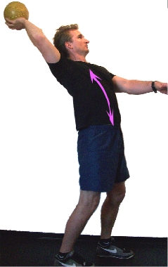
Traditional anatomy teaches that the erector spinae group of muscles helps straighten the spine into extension and keep it erect over the pelvis (Gray 1995). That’s why we typically give clients spine extension exercises such as “Supermans” or repetitions on the back extension machine. We are also taught that the rectus abdominis helps flex the spine (Golding & Golding 2003), and therefore we recommend that clients do sit-ups. However, the real-life function of the erector spinae muscles is to slow down forward flexion of the spine as we bend forward, as when reaching over to pick something up (Price 2008). Conversely, the real-life function of the rectus abdominis is to slow down trunk extension, as when we reach overhead to wash our hair.
Erector spinae muscles that are able to lengthen under load effectively slow the spine as it bends forward. They can also generate tension as they lengthen, which can be used to pull the spine back up into an upright position. The abdominals function as a counterpart to the erector spinae group by lengthening under load to ensure that the spine does not extend too far back as the torso is pulled upright. When these muscles are healthy and functional, they work together like alternate sets of bungee cords to prevent the spine from arching or rounding too far or too quickly, thereby minimizing stress to the spine and torso.
As such, spinal extension and abdominal crunches are not the best ways to strengthen the back and abdominal muscles. A more effective exercise would be to have a client bend down and pick up a medicine ball and then reach back overhead and throw it to you. This exercise requires the erectors to lengthen in order to slow down the spine as it bends forward so the client can pick up the ball, and it requires the abdominals to lengthen in order to slow down the spine as it arches backward as the client prepares to throw the ball (see Figures 3a & 3b).
Example 4: The Hip Flexors

The hip flexor muscles originate from the lumbar spine (the psoas major and minor) and pelvis (the iliacus) and attach to the top of the femur (Golding & Golding 2003). According to traditional anatomy texts, the hip flexors bring the leg and spine toward each other and externally rotate the leg (Gray 1995). However, in real life the hip flexors help keep the spine balanced over the lower body during activities like walking and running. The presence of gravity and ground reaction forces means that the function of the hip flexors is actually to slow down hip/leg extension as the trail leg travels behind the body when walking, running, lunging and so on. The lengthening of these muscles also creates bungee cord–like tension, which—when released—enables the leg to swing through and under the pelvis with ease (hip flexion).
As a result of our modern-day environment, which involves many hours of sitting, the hip flexors can become chronically shortened (McGill 2002). Naturally, this causes problems when a person tries to walk or run, as the hip flexor muscles can no longer lengthen effectively. Therefore, it is important to keep these muscles supple and strong so they can work in a lengthening manner to assist with locomotion. Instead of doing leg lifts to strengthen the hip flexors, a better choice would be to add reverse lunges to training programs (or simply to coach clients in stepping backward) to practice extending the hips (see Figure 4). This kind of exercise will push the hip/leg complex into extension and train the hip flexors how to lengthen under load, thus slowing down hip/leg extension.
A Fresh Approach to Anatomy and Exercise
By understanding anatomy in real-life terms and thinking about how muscles slow down the forces of nature by lengthening like bungee cords, you can design more effective exercises to build strength, improve function and help eliminate pain. A keen understanding of individual muscles and how they connect through fascial lines leads to integrated, whole-body program design. The result: fun and highly effective moves that help clients reach their health and fitness goals faster and more safely.
Source: Burke et. al. 2001.
video web extra
@VIDEO_FILE:/files/video/201009-ifj-01.mp4:500:281@
@VIDEO_FILE:/files/video/201009-ifj-02.mp4:500:281@
@VIDEO_FILE:/files/video/201009-ifj-03.mp4:500:281@