Total Joint Replacement: Knee and Hip
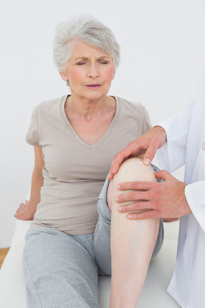
Mr. Brown is a 68-year-old retired postal worker who stays active with golf and tennis, but he complains of severe pain and swelling in his left knee, which he cannot straighten completely. The pain limits his ability to do the things he loves, but he is otherwise comfortable during daily activities. Based on X-rays and a clinical exam, Mr. Brown has symptomatic knee osteoarthritis.
The initial plan of action includes activity modification, an ambulatory device (cane) as needed, over-the-counter medications and an exercise program. A year later, Mr. Brown’s pain is not only limiting his ability to participate in his recreational activities; it is also disturbing his sleep and hindering some daily activities, such as climbing stairs. As a result, he is scheduled for a total knee replacement, or knee arthroplasty.
Mrs. Kramer is a 57-year-old hair stylist who enjoys taking fitness classes. She has always been in great shape, but recently she’s been unable to participate in her exercise routine because
of right hip pain. She used to be a long-distance runner, but she stopped running regularly about 10 years ago. Today, she describes having pain in her groin that gets worse with prolonged standing and walking. Her hip has become stiff, and she lacks the flexibility she once had.
Mrs. Kramer’s pain makes it difficult for her to get through a full workday. She has tried a course of physical therapy, and over-the-counter medications are no longer helping. An X-ray and clinical exam show that Mrs. Kramer has symptomatic hip osteoarthritis. Given that she can’t tolerate her daily activities, and conservative management has failed, she is scheduled for a right hip replacement, posterior approach.
If you’ve worked with a client like Mr. Brown or Mrs.
Kramer, you know how frustrating and limiting osteoarthritis pain can be. When it leads to the need for total joint replacement, it’s vital that fitness professionals understand the process and how to work with allied health professionals to help the client get back to an active life. This article reviews OA, examines the steps involved in total knee and hip replacement, and explains how fitness professionals fit into the treatment and recovery plan.
Osteoarthritis Review
Osteoarthritis is a degenerative disease of the synovial joints that results in progressive articular cartilage loss and pain. It is the most common joint disorder in the United States, with symptomatic OA occurring in 10% of men and 13% of women aged 60 or older (Zhang & Jordan 2010). While certain risk factors—such as age, gender and genetics— cannot be changed, multiple risk factors can be adjusted to reduce the likelihood of suffering from symptomatic OA. Modifiable risk factors include obesity, trauma and muscle weakness. The rising incidence of obesity indicates that OA rates are likely to increase (Muthuri et al. 2011; Christensen et al. 2005).
On X-rays, individuals with OA may be asymptomatic. If they are symptomatic, they suffer from joint pain, swelling, stiffness and reduced range of motion in the affected joint. Some develop deformity or joint malalignment. X-rays are best taken in a standing, or weight-bearing, position. Clinicians often note a narrowing of the joint space, osteophytes (bone spurs) and other, subtler changes to the bony architecture.
Note: The following information is for general education purposes only. Seek further continuing education on the topic of total joint replacement before designing a program for a client, and understand the steps involved in working in concert with physical therapists and physicians.
Conservative Knee Osteoarthritis Management
Usually, physicians will initiate a conservative, or nonoperative, treatment plan for OA. Measures include over-the-counter anti-inflammatory medications, weight loss programs and physical therapy or exercise. A combination of supervised exercises and a home program has shown the best results and is supported by the American Academy of Orthopaedic Surgeons (AAOS 2013; Lorig et al. 1985; Coleman et al. 2012). This course of action is likely to involve fitness professionals who understand the special needs of clients with OA and who stay within scope of practice while working with allied health professionals.
In certain situations, doctors may prescribe a brace. Bracing may be useful if the osteoarthritic disease is focused to one side of the knee (medial or lateral), also known as unicompartmental disease. A custom-fit brace may help unload the resulting varus (knock-kneed) or valgus (bowlegged) deformity created by unicompartmental disease (Kirkley et al. 1999).
The AAOS does not recommend the use of viscoelastic injections, and reports that the evidence for using corticosteroid injections is inconclusive (AAOS does not weigh in for or against their use) (AAOS 2013).
Knee Osteoarthritis
Operative Management
In past years, arthroscopic partial meniscectomy (surgical removal of the meniscus) has been favored for symptomatic OA, as well as for a torn meniscus; however, recent evidence argues against this recommendation (AAOS 2013). Beyond meniscectomies, nonarthroplasty options—which include high tibial osteotomy (surgical removal of a wedge of the tibia, in order to straighten the leg)—are common for younger adults. Nonarthroplasty procedures delay the inevitable arthroplasty, which is the ultimate solution. A delay avoids the need for a revision arthroplasty, since total joint replacement has a lifespan of approximately 15–30 years.
Anatomy Review: Knee Surgery
The medial parapatellar surgical approach is standard for total knee arthroplasty. In this approach, the incision creates an inter-muscular plane between the rectus femoris and the vastus medialis, beginning approximately 5 centimeters above the superior pole of the patella and extending to the level of the tibial tubercle. The surgeon then dissects down the subcutaneous tissues and divides the vastus medialis and quadriceps tendon, exposing the knee joint. Surgeons are careful to avoid injuring the superior lateral genicular artery (at risk when dissection occurs along the lateral aspect of the knee joint) or the infrapatellar branch of the saphenous nerve (found medially). The superior lateral genicular artery provides an important blood supply to the joint, while injury to the branch of the saphenous nerve would result in a postoperative neuroma. The overall incidence of total knee arthroplasty complications is 1% (Parvizi et al. 2007).
Total Knee Arthroplasty
Postoperative Course
Surgery generally takes about 2 hours, after which the patient travels to a recovery unit. Once stable, he may transfer to a general hospital floor. The hospital stay is commonly 2–3 nights, with physical therapy once or twice a day. The ROM goal during inpatient care ranges from approximately -10 degrees (extension) to 80 degrees (flexion). Those with preexisting stiffness
may have modified goal ranges. A continuous passive motion machine—a motorized device the patient dons to do gentle flexion and extension ROM while lying in bed—may be used postoperatively. The data on CPM devices is controversial, and implementation is generally based on the surgeon’s preference (Harvey, Brosseau & Herbert 2014).
Inpatient therapeutic exercises may include the following:
- bed mobility and transfers
- ambulation with an assistive device (crutches or walker)
- ROM: flexion and extension (therapist may help with passive ROM)
- heel slides (active ROM)
- isometric quad sets, hamstring sets and gluteal sets
- straight-leg raises
Patients are then discharged to home physical therapy or discharged to a rehabilitation facility for a short-term stay. To go home, they must demonstrate that their home is safe, and must show they can effectively and safely handle the activities of daily living.
Outpatient Physical Therapy
The goal of the next phase focuses on motion, strength, endurance and balance (Lin et al. 2009; Fitzgerald et al. 2011).
RANGE OF MOTION
By the sixth week after the operation, the knee should be able to range from 0 degrees of extension to 110 degrees of flexion. Passive and active exercises help to increase motion. The stationary bike is a great tool for improving ROM; exercise should begin with partial revolutions and then progress to full revolutions as tolerated. Initially there should be no resistance.
STRENGTH
Muscle strengthening focuses on the knee extensors (rectus femoris, vastus lateralis, vastus medialis and vastus intermedius) and flexors (biceps femoris, semitendinosus and semi-
membranosus). After any operation, however, it’s important to strengthen the upper extremities, the contralateral lower extremity and the core. Fitness professionals who are working with physicians and physical therapists should inquire about this program design approach for the client in question.
Strengthening exercises for outpatient physical therapy focus on these elements:
- progressing beyond isometric quadriceps, hamstring and
gluteal exercises - straight-leg raises in four planes (flexion, abduction, adduc-
tion and extension) - bridging
- sit-to-stand movements
- balance activities
- a pool program (after the wound is healed)
EXERCISES
Four-Way Straight-Leg Raises
- Lie on back, unaffected knee bent, foot flat. *Engage core, and lift affected leg ~12 inches from floor, ankle dorsiflexed. Hold 5 seconds, and slowly lower leg to floor. Do 10 reps, 3 times per day.*
- Roll to side and repeat asterisked instructions above.
- Roll onto stomach, placing hands comfortably under fore-
head, and repeat asterisked instructions above. - Roll to other side, placing lower leg in comfortably bent
position, and repeat asterisked instructions above.
Bridging
- Lie on back, hands resting by sides, knees bent, feet flat on floor.
- Keep feet under knees, tighten core muscles, and elevate hips.
- Pause when straight line is formed from shoulders to knees.
- Hold 5 seconds and slowly lower back down.
- Do 10 reps, 3 times per day.
Sit to Stand
- Sit in chair with good, upright posture.
- Place feet under knees, hands on lap.
- Tighten core, and push up through heels to standing position.
- Pause briefly, then slowly lower back down to chair.
- Do 10 reps, 3 times per day.
BALANCE ACTIVITIES
Proprioceptive training improves body/spatial awareness of the affected extremity in functional activities. The following progression is an example:
- Begin standing, feet hip-width apart.
- Lift one leg off ground, knee slightly bent.
- Maintain balance for up to 15 seconds.
- Repeat with other leg.
- If client can maintain balance for 15 seconds without difficulty, add challenges such as reaching for object in front, across body, overhead and, finally, on floor.
ENDURANCE
The purpose of endurance training is to increase overall cardiovascular fitness, which may be significantly reduced following arthroscopy. Many people are in severe pain before surgery and, therefore, have difficulty exercising. It is not unusual for them to be quite deconditioned.
A client is generally ready to graduate from physical therapy when he can demonstrate the following:
- a smooth, independent gait
- pain-free ROM
- full, functional ROM
- overall strength graded at 4+/5 or better
- age-appropriate balance skills
Total Knee Arthroplasty Postrehabilitation
Postrehab goals include a return to recreational sports or activities, as well as strength, endurance and proprioception enhancement. The focus is on maintaining good quadriceps and hamstring strength. Below is a list of recommended post-rehab exercises:
Step-Downs
- Stand tall on 12-inch step with feet hip-width apart.
- Leading with unaffected extremity, step down onto floor.
- Gently tap floor with leading heel, then push through leg on
step, rising back to start position. - Do 3 sets of 10 reps per side.
Variation: Hold weights while stepping, or toss medicine ball while stepping down.
BOSU® Balance Trainer Bridge
- Perform as described above, but instead of placing feet flat
on mat, place feet on rounded side of BOSU ball. Repeat 3 sets of 10 reps per side.
Progression: Place feet on flat surface of BOSU ball.
TheraBand™ Walks
- Place TheraBand loop around ankles and perform side steps for 25 feet in one direction.
- Switch directions and repeat.
- Perform 3 sets in each direction.
Progression: Add side squats.
Single-Leg Stance
- Perform single-leg stance (on affected extremity) in front of
mirror. - Hold steady for 15 seconds before progressing.
Progression: Do single-leg stance while tossing ball (small or large) or while reaching for objects away from body.
Lower-Extremity Flexibility
Continue a generalized flexibility program. Place special attention on calf and hamstring flexibility, as these muscle groups tend to cramp and get tight during rehab progression.
Conservative Hip Osteoarthritis Management
When possible, physicians treat people with symptomatic hip OA through conservative means before opting for operative management. Over-the-counter anti-inflammatories, weight loss programs and physical therapy or exercise all play an important role.
Hip Osteoarthritis Operative Management
In past years, hip resurfacing, an alternative to total hip arthroplasty, has been available to a subset of individuals. In this procedure, doctors place a metal cap over the head of the femur, and this cap articulates with a metal cup placed in the acetabulum, or pelvis socket. The proposed advantages are a lower dislocation rate and faster recovery. However, certain criteria, including good proximal bone stock, must be met. The operating surgeon determines whether or not an individual qualifies for this procedure. More commonly, total hip arthroplasty is recommended and performed as the definitive treatment for symptomatic hip OA.
Anatomy Review: Hip Surgery
There are two standard surgical approaches for total hip arthroplasty: anterior and posterior. Both have a long, successful track record, and the surgeon generally determines which one to use. In the postoperative setting, the choice is an important factor, as it influences precautions and mobility options (Hoppenfeld, deBoer & Buckley 2009).
The posterior approach utilizes an intermuscular plane, splitting the gluteus maximus. During the dissection down to the hip capsule, the surgeon must be careful to avoid injuring the sciatic nerve, which most commonly travels below the piriformis. Postoperatively, people who have undergone a total hip arthroplasty are told by their surgeons to avoid certain motions in order to reduce their risk of dislocation. With the posterior approach, one must avoid hip flexion, internal rotation and adduction (e.g., sitting cross-legged) (Hoppenfeld, deBoer & Buckley 2009).
The plane of dissection for the anterior approach is internervous, between the sartorius and the tensor fasciae latae. The surgeon must carefully avoid the lateral femoral cutaneous nerve, which carries sensation to the anterior, proximal thigh. Postoperatively, people who have undergone the anterior approach must avoid hip extension and external rotation (e.g., getting out of a deep seat in a car) (Hoppenfeld, deBoer & Buckley 2009).
Total Hip Arthroplasty Postoperative Course
Surgery generally takes about 2 hours, after which the patient travels to a recovery unit. Once stable, she transfers to a general hospital floor. The hospital stay is commonly 2–3 nights, with physical therapy sessions occurring once or twice a day.
Inpatient therapeutic exercises include the following:
- bed mobility and transfers
- ambulation with an assistive device (crutches or walker)
- long-arc quad sets (seated knee extensions without resis-
tance) - isometric quad sets, hamstring sets and gluteal sets
- straight-leg raises
As with knee procedures, patients must be cleared for safe discharge to home by a therapist before they leave the hospital. Some people may need a short stay in rehab to reach this goal.
Outpatient Physical Therapy
The next phase focuses on improving motion, strength, endurance and balance.
RANGE OF MOTION
By the sixth week after the operation, individuals should be able to comfortably flex their hip to 90 degrees and abduct to 30 degrees.
STRENGTH
An effective muscle-strengthening program addresses the entire hip girdle, but particular attention must be paid to the hip abductor and extensor muscle groups. To review the anatomy: The gluteus maximus is the primary hip extensor, while the gluteus medius is a primary abductor. The psoas and quadriceps work together to perform hip flexion, and the adductor group approximates the femur toward the midline. Balancing effort among the hip abductors, adductors, flexors and extensors is essential to establish a smooth, comfortable gait. It’s also important to focus on core strength and overall contralateral lower-extremity strength.
Strengthening exercises for outpatient physical therapy focus on these elements:
- standing hip abduction
- wall sits
- side squats
- balance activities
- a pool program (once the wound is healed)
The goal is to progress beyond isometric quadriceps, hamstring and gluteal exercises.
EXERCISES
Standing Hip Abduction
- Begin standing tall on unaffected leg, core engaged, feet hip-width apart. Swing affected leg (with control) out to ~30 degrees of abduction while using upper extremities to assist balance. Do 3 sets of 10 reps per side.
Wall Slides
- Stand against wall, feet ~24 inches away. Slowly lower toward 90 degrees of hip flexion, pause, and rise back up. Keep knees pointed forward, and do not cross toes. Do 3 sets of 10 reps.
Side Squats
- Stand tall, feet hip-width apart. With knees pointing forward, step out to one side ~24 inches. Lower into squat, maintaining appropriate form. Return to start. Do 3 sets of 10 reps per side.
Progression: Add resistance with tubing or pulleys.
BALANCE ACTIVITIES
See balance activities for knee rehab. Progress the single-leg stance exercises from standing on one leg to incorporating distracting activities.
ENDURANCE
The aim here is to improve endurance with treadmill workouts and pool walking. Cycling is an option, but the affected hip must not flex more than 90 degrees if the surgeon used a posterior approach. A client is generally ready to graduate from physical therapy when she can demonstrate the following:
- a smooth, independent gait
- pain-free ROM
- full, functional ROM
- overall strength graded at 4+/5 or better
- age-appropriate balance skills
Postrehab: Total Hip Arthroplasty
Postrehab goals include a return to recreational sports or activities, as well as improvements in strength, endurance and proprioception. The focus is on maintaining good hip extensor and hip abductor strength. Below is a list of recommended postrehab exercises:
Step-Ups
- Stand tall, facing 12-inch step, feet hip-width apart. Leading with affected extremity, step up onto step, foot flat. Raise body upward until standing tall on step. Pause, then slowly lower back down. Do 3 sets of 10 reps per side.
Progression: Step onto more challenging surface, such as
BOSU ball.
Walking Lunges
- Perform walking lunges with appropriate form for 25 feet. Repeat 3 times.
Progression: Hold weights or medicine ball while lunging, or
perform lunges on grass (more challenging surface).
Wall Sits With Stability Ball
- Stand against wall, feet ~24 inches in front, with large therapeutic ball placed in small of back. Slide down to squat position; hold for 30 seconds. Push through heels and return to standing. Do 3–5 reps.
Progression: From squat position, toss medicine ball or do single-leg extension.
Single-Leg Stance
- Stand on one leg (on affected extremity) in front of mirror.
Hold steady for 15 seconds before progressing.
Progression: Do single-leg stance with ball toss (small or large ball), or reach for object away from body.
Lower-Extremity Flexibility
- Continue a generalized flexibility program. Place special attention on calf and hamstrings flexibility, as these muscle groups tend to cramp and get tight during rehab progression.
POSTOPERATIVE PRECAUTIONS
FOR TOTAL HIP ARTHROPLASTY
Follow all precautions for at least 3 months or as directed by the surgeon.
Posterior dislocation: no hip flexion greater than 90 degrees, hip internal rotation or adduction beyond neutral.
Anterior dislocation: no hip extension or hip external rotation beyond neutral. Note: Avoid bridging or prone exercises.
Life After Surgery
What happened to Mr. Brown and Mrs. Kramer? Following the surgery, Mr. Brown recovered well in the hospital and participated in physical therapy. After graduating from physical therapy, he began a postrehab program with a personal trainer at his local fitness facility. The next spring, he returned to golf and doubles tennis.
Following her total hip replacement, Mrs. Kramer recovered well in the hospital and participated in a course of physical therapy. After graduating, she joined a postrehab program at her local gym with a personal trainer. The trainer was careful to observe posterior-dislocation precautions until the surgeon gave the green light to proceed. Mrs. Kramer resumed her fitness routine postoperatively.
With the right knowledge, dedication to working with allied health professionals, patience, and respect for postoperative precautions, fitness professionals are primed to help clients who have had a total joint replacement not only recover but thrive and enjoy additional years of inspirational movement.